 Dr. T.B.S. Buxi
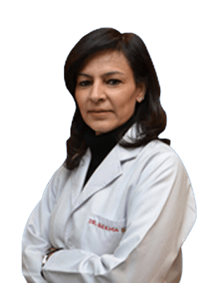 Dr. Seema Sud
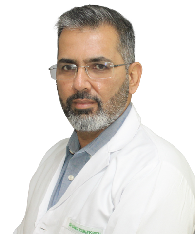 Dr. Samarjit S Ghuman
 Dr. Kishan S Rawat
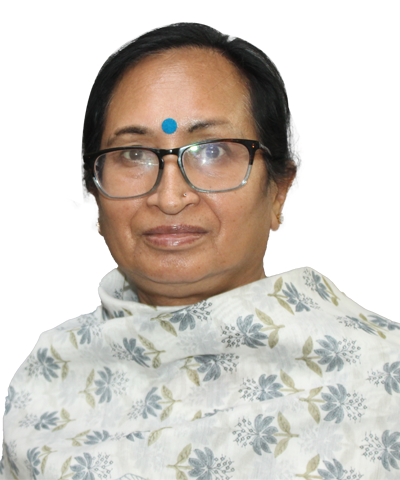 Dr. Sushma Saroa
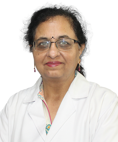 Dr. Anurag Yadav
MRI also known as Nuclear Magnetic Resonance Imaging (NMRI), is a technique used in radiology to visualize in detail the internal structures of the body. It provides excellent contrast among various soft tissues of the body, and is very useful in imaging the brain, nerves, spine, joints, bones, muscles, blood vessels, chest, breast, heart, abdomen and pelvis including the rectum, prostate, uterus and ovaries. Compared with other medical imaging techniques, it does not use X-ray radiation and is therefore safe during pregnancy and in the newborns and children.
We have a state-of-the-art 3Tesla Siemens MRI machine with an open-bore magnet, the fi rst of its kind in India. This results in fast scanning and causes less claustrophobia. In addition to the routine T1W and T2W scans performed for stroke, blood clots, infarcts, tumours and infections, we do high-end specialized MR studies such as MR spectroscopy, functional imaging, perfusion and diffusion studies, post-gadolinium contrast studies, MRI brain with epilepsy protocol, angiography and venography. Our centre is a referral base for patients not only from New Delhi (NCR), but from other parts of India and abroad.
Whole-spine screening is possible in patients undergoing MR for disc prolapse, sciatica, back pain and cancers.
A wide range of other advanced MRI studies include MR cholangiopancreatography (especially for liver donors and patients with gallstones), MR urography, MR Defaecography, MR Enterography, MR fi stulography in anal fi stulae, evaluation of the breasts by MR mammography, prostate MRI by dynamic contrast, MR diffusion and spectroscopy. Dedicated studies are performed for patients with deafness for cochlear implants. We also perform high-resolution cardiac MRI for heart attacks, tumours, muscle pathologies and congenital heart diseases.
We undertake renal angiography, peripheral angiography, triphasic angiography for the liver, whole-body MRI, whole-body angiography, wholebody diffusion for cancer spread.
2D and 3D reconstructions, dynamic MR and MR arthrography of all joints including temporomandibular, shoulder, elbow, wrist, hand, hip, sacroiliac, knee, ankle, foot and atlantoaxial can be obtained using high-resolution MR techniques
Computed tomography (CT) or computed axial tomography (CAT) is an imaging method using focused X-ray beams to generate an image of a body part in an axial plane to acquire 3D data. The Department of CT Scan at SGRH is accredited with many fi rsts: It was the fi rst to install the whole-body CT in 1983, the fi rst spiral CT in 2000, the fi rst multidetector CT in 2004 in North India, and now the low-dose CT in 2011 – the fi rst in south-east Asia
The 128-slice low-dose CT is the latest in CT technology. Radiation, even in diagnostic imaging, potentially increases the patient’s risk to develop cancer later in life. The low-dose CT at our centre reduces the radiation per study by up to 40% without compromising the image quality. The iDose technology of Philips is being used in every scan to tailor the reduction in radiation dose.
All CT scan studies are performed with specialized procedures such as CT angiographies – whole-body, coronary, peripheral, pulmonary and abdominal angiographies; liver, kidney, small bowel transplant work-ups; imaging of congenital heart diseases; temporal bone imaging, dentascan; oncology imaging; CT enterography; non-invasive CT bronchoscopy and colonoscopy; 3D reconstruction of joints and musculoskeletal system in tumours and trauma; CTguided FNACs, biopsies and drain placements; sterotactic biopsies; CT brain perfusion studies are done in routine practice. Newer software for assessment of emphysema for lung volume reduction surgeries, computer-aided detection of lung nodule assessment and aortic valve sizing for TAVI have also been added.
Each patient is evaluated by a full-time anaesthesiologist for safety of contrast administration and providing anaesthesia/sedation whenever required. There are extensive discussions with the referring clinicians before and after the scans, and all scans are interpreted by highly qualifi ed, well-trained, experienced radiologists taking into account the clinical findings.
The Department of CT Scan is recognized, as an integral part of the hospital, for DNB postgraduate training in radiodiagnosis. A number of research projects, fellowships and onsite training programmes are always ongoing. The department is accredited with NABH, NABL, and meets all the patient safety standards.
CT scan – Room no: 21, MRI – Room no: 22, Ground Floor, Old Building, Ext: 1912/1909